The healing of skin wounds provides a classical example of the combination of regeneration and repair. Wound healing can be accomplished in one of the following two ways:
(i) Healing by first intention (ii) Healing by second intention
Table of Contents
(i) Healing by first intention:
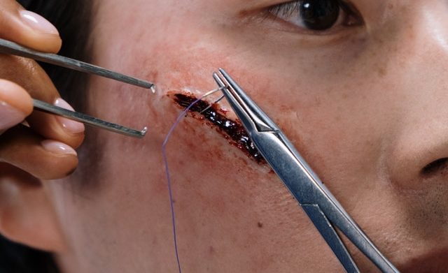
One of the simplest examples of wound repair is the healing of a clean, uninfected surgical incision approximated by surgical sutures. This is referred to as healing by the first intention. The incision causes only focal disruption of epithelial basement membrane continuity and death or a relatively few epithelial and connective tissue cells.
As a result, epithelial regeneration predominates over fibrosis. The narrow incisional space rapidly fills with fibrin-clotted blood; dehydration at the surface produces a scar to cover and protect the healing repair site.
On day first, neutrophils are seen at the incision margin, migrating toward the fibrin clot. Basal cells at the cut edge of the epidermis begin to exhibit increased mitotic activity. Within 24 to 48 hours, epithelial cells from both edges have begun to migrate and proliferate along with the dermis, depositing basement membrane components as they progress. The cells meet in the midline beneath the surface scab, yielding the thin but continuous epithelial layer.
On days two to three neutrophils have been largely replaced by macrophages, and granulation tissue progressively invades the incision space. Collagen fibers are now evident at the incision margins, but these are vertically oriented and do not bridge the incision. Epithelial cell proliferation continues, yielding a thickened epidermal covering layer.
On days 4 to 5 neovascularization reaches its peak as granulation tissue fills the incisional space. Collagen fibrils become more abundant and begin to bridge the incision. The epidermis recovers its normal thickness as differentiation of surface cells yields a mature epidermal architecture with surface keratinization.
In the second week, there is continued collagen accumulation and fibroblast proliferation. The leukocyte infiltrates, edema, and increased vascularity are substantially diminished. The long process of blanching begins, accomplished by increasing collagen deposition within the incisional scar and the regression of vascular channels.
In the first month, the scar comprises a cellular connective tissue largely devoid of inflammatory cells and covered by an essentially normal epidermis. However, the dermal appendages were destroyed in the line of the incision and permanently lost.
(ii) Healing by second intention:
When cell or tissue loss is more extensive, as in infractions, inflammatory ulceration, abscess formation, or even just large wounds, the reparative process is more complex. In these situations, regeneration of parenchymal cells alone cannot restore the original architecture. As a result, there is extensive ingrowth of granulation tissue from the wound margin, followed in time by the accumulation of extracellular matrix and scarring. This form of healing is referred to as secondary union or healing by the second intention.
Phases of Wound Healing
The response of tissue to injury goes through several phases which are:
(a) Inflammatory phase
(b) Proliferative phase
(c) Maturation phase
(a) Inflammatory Phase:
Also called the lag or executive phase, the inflammatory phase is characterized by vascular and cellular responses that occur immediately after tissue injury takes place. The length of this phase lasts for about 1-4 days. The following events occur during this phase:
1. Blood clot formation:
Right after tissue injury takes place, vasoconstriction of vessels occurs, and in an attempt to stop or control bleeding, a fibrinoplatelet clot forms. Lasting for about 5 to 10 minutes, this reaction is followed by vasodilation of the venules. Vasoconstriction is stopped as norepinephrine is destroyed by the intracellular enzymes. The result is increased permeability of the capillary due to the destruction of norepinephrine and the release of histamine.
2. Wound becomes edematous:
Damage of the microcirculation results to the infiltration of blood elements such as antibodies, plasma proteins, electrolytes, complement, and water for approximately 2 to 3 days after the tissue injury. This mechanism causes the occurrence of edema, warmth, redness, and pain on the affected area.
3. Phagocytes engulf debris of damaged tissue and blood clot:
The first leukocytes that move into the damaged tissue are the neutrophils. The component of WBC that transforms to macrophages to engulf the debris, monocytes, transports the phagocytized debris from the injured area. Antigen antibodies also appear and the basal cells present at the edges of the wound undergo mitosis and the resulting daughter cells migrate. Through this activity, the secretion of proteolytic enzymes and their breakdown at the base of the clot is made possible. Thus, the break in the continuity of the cells is progressively bridged and by about 24 to 48 hours the sides of the wound would eventually meet. Hyperplastic bone marrow activity enhances cell migration progressing to the next phase of wound healing.
(b) Proliferative Phase:
The proliferative phase also called the fibroblastic or connective tissue phase, is the time where fibroblasts are multiplying and a lattice framework is formed for migrating cells. The proliferative phase occurs during the 5th to the 20th day after tissue injury took place. The following events are noted during this phase of wound healing:
1. Granulation tissue forms:
By this time, epithelial cells form beds at the edges of the wound. These beds are the ones that develop into capillaries. These capillaries serve as the nutritional source for the new granulation tissue.
2. Collagen production:
The primary component of replaced connective tissue is collagen. It is the fibroblasts that initiate the synthesis of collagen and mucopolysaccharides. Chains of amino acids convert into fibers of increasing length and diameter in about 2 to 4 weeks. The formed fibers become a well-structured pattern of packed bundles. Collagen synthesis causes depletion in the number of capillaries.
(c) Increased wound tensile strength:
Synthesis of collagen and lysis of capillaries results in the increased tensile strength of the wound. About 3% to 5% of the original skin strength is the amount of skin present 2 weeks after the injury. About in a month, only 35% to 59% of wound strength has been reached. Nevermore than 70% to 80% of the strength is regained after the wound has occurred. Vitamin C aid in the metabolic process necessary for wound healing.
Also read: What are Inflammatory Mediators