
Table of Contents
Cardiovascular system
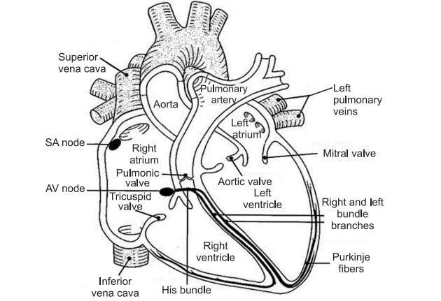
The Conduction System of the Heart: The heart is a muscular organ the size of a closed fist that is positioned in the chest behind the sternum and above the diaphragm, between the lungs. It is encased in the pericardium. The aorta, pulmonary arteries and veins, and the vena cava are all connected to the base of the heart on its superior end. The apex, or inferior tip of the heart, is located slightly above the diaphragm. The heart’s base runs parallel to the body’s midline, with the apex pointing to the left. Because the heart is angled to the left, around two-thirds of the heart’s mass is located on the left side of the body, while the remaining one-third is located on the right.
The heart is the pump that is responsible for maintaining adequate circulation of oxygenated blood around the vascular network of the body. It takes in deoxygenated blood through the veins and delivers it to the lungs for oxygenation before pumping it into the various arteries.
Anatomy of the Heart
The heart is contained within a fluid-filled chamber known as the pericardium. The pericardium is a unique membrane that lines and protects the pericardial cavity’s walls and lining. The pericardium is a type of serous membrane that produces serous fluid to lubricate and protect the heart and its surrounding organs from friction. The pericardium holds the heart in place and maintains a hollow space for the heart to expand when it is full, in addition to providing lubrication. The pericardium is divided into two layers: a visceral layer that covers the outside of the heart and a parietal layer that creates a sac around the pericardial cavity’s perimeter. Epicardium, myocardium, and endocardium are the three layers that make up the heart wall.
Epicardium: The epicardium is the outermost layer of the heart wall. It is also referred to as visceral pericardium, which is the inner layer of the pericardium. The epicardium is a thin layer of serous membrane that helps to lubricate and protect the outside of the heart.
Myocardium: The myocardium is the thick middle layer of the heart wall and consists of numerous layers of cardiac muscle fibers that wrap around the heart. Contraction of the myocardium pumps blood out of the heart into the aorta and pulmonary trunk arteries.
Endocardium: The endocardium is the simple squamous endothelium layer that lines inside the heart. The endocardium is very smooth and is responsible for keeping blood from sticking to the inside of the heart and forming potentially deadly blood clots. The thickness of the heart wall varies in different parts of the heart. The atria of the heart have a very thin myocardium because they do not need to pump blood very far, but only to the nearby ventricles. The ventricles, on other hand, have a very thick myocardium to pump blood to the lungs or throughout the entire body. The right side of the heart has less myocardium in its walls than the left side because the left side has to pump blood through the entire body while the right side only has to pump to the lungs.
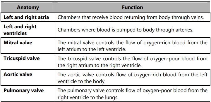
Heart Chambers: The heart contains four chambers: the right atrium, left atrium, right ventricle, and, left ventricle. The atria are smaller than the ventricles and have thinner, less muscular walls than the ventricles. The atria act as receiving chambers for blood, so they are connected to the veins that carry blood to the heart. The ventricles are the larger, stronger pumping chambers that send blood out of the heart. The ventricles are connected to the arteries that carry blood away from the heart.
Heart Valves: The heart functions by pumping blood both to the lungs and to the systems of the body. To prevent blood from flowing backward or “regurgitating” back into the heart, a system of one-way valves are present in the heart. The heart valves can be divided into two types: atrioventricular and semilunar valves.
Atrioventricular Valves (AV): The AV valves are located in the middle of the heart between the atria and ventricles and only allow blood to flow from the atria into the ventricles. The AV valve on the right side of the heart is called the tricuspid valve because it is made of three cusps (flaps) that separate to allow blood to pass through and flow backward. The AV valve on the left side of the heart is called the mitral valve or the bicuspid valve because it has two cusps. The AV valves are attached on the ventricular side to tough strings called chordae tendineae. The chordae tendineae pull on the AV valves to keep them from folding backward and allowing blood to flow backward.
Semilunar Valves: The semilunar valves, so named for the crescent moon shape of their cusps, are located between the ventricles and the arteries that carry blood away from the heart. The semilunar valve on the right side of the heart is the pulmonary valve, so named because it prevents the backflow of blood from the pulmonary trunk into the right ventricle. The semilunar valve on the left side of the heart is the aortic valve, named for the fact that it prevents the aorta from flowing blood back into the left ventricle. The semilunar valves are smaller than the AV valves and do not have chordae tendineae to hold them in place. Instead, the cusps of the semilunar valves are cup-shaped to catch the backward flowing blood and use the blood pressure to snap shut.
Conduction System of the Heart
The heart has the ability to create its own rhythm as well as transmit the signals required to maintain and coordinate that rhythm throughout its various structures. The conduction system, which sets the pace for the rest of the cardiac muscle cells, is formed by around 1% of the cardiac muscle cells in the heart.
The conduction system begins with the heart’s pacemaker and the sinoatrial (SA) node, a tiny bundle of cells. The SA node is found in the right atrium’s wall, below the superior vena cava. The SA node is in charge of regulating the heart’s overall rhythm and directly instructs the atria to contract. The signal from the SA node is picked up by the atrioventricular (AV) node, which is a mass of conductive tissue.
The AV node is positioned in the inferior section of the interatrial septum in the right atrium. The signal sent by the SA node is picked up by the AV node and transmitted to the bundle of His. The AV bundle is a conductive tissue strand that goes from the interventricular septum to the interatrial septum. In the interventricular septum, the AV bundle splits into left and right branches, which continue flowing through the septum until they reach the apex of the heart. There are numerous bundle branches on the left and right. Purkinje fibers, which transport the signal to the ventricle walls, cause cardiac muscle cells to contract in a coordinated manner, allowing blood to be efficiently pumped out of the heart.
Also read: What are Inflammatory Mediators